Principles of origami used to position sensors inside 3D-bioprinted tissues
Innovative platform from TAU combines science with art
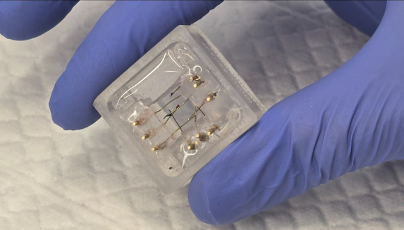
Support this researchResearchers at Tel Aviv University (TAU) relied on principles of origami, the Japanese art of paperfolding, to position sensors inside 3D-bioprinted tissue models. Instead of bioprinting tissue over the sensors, which proved to be impractical, they designed and produced an origami-inspired structure that folds around the fabricated tissue, allowing the insertion of sensors into precisely pre-defined locations.
The study was a joint effort of researchers from several units at TAU: the School of Neurobiology, Biochemistry and Biophysics, the Koum Center for Nanoscience and Nanotechnology, the Department of Biomedical Engineering, the Sagol Center for Regenerative Medicine, the Sagol School of Neuroscience, and the Drimmer-Fischler Family Stem Cell Core Laboratory for Regenerative Medicine. The research was led by Professor Ben Maoz and Professor Uri Ashery and was published on April 18, 2024, in Advanced Science.
“The use of 3D-bioprinters to print biological tissue models for research is already widespread,” Professor Maoz explains. “In existing technologies, the printer head moves back and forth, printing layer upon layer of the required tissue. This method, however, has a significant drawback: The tissue cannot be bioprinted over a set of sensors needed to provide information about its inner cells, because in the process of printing the printer head breaks the sensors. We propose a new approach to the complex problem: origami.”
The innovation is based on an original synergy between science with art. Using Computer Aided Design software, the researchers were inspired by origami paper-folding to design a multi-sensing structure customized for a specific tissue model. This structure incorporates various sensors for monitoring the electrical activity or resistance of cells in precisely chosen locations within the tissue. The computer model is used to manufacture a physical structure which is then folded around the bioprinted tissue so that each sensor is inserted into its predefined position inside the tissue. The TAU team has named their novel platform the “Multi-Sensor Origami Platform,” or MSOP.
The new method’s effectiveness was demonstrated on 3D-bioprinted brain tissues, with the inserted sensors recording neuronal electrical activity. The researchers emphasize, however, that the system is both modular and versatile: It can place any number and any type of sensors in any chosen position within any type of 3D-bioprinted tissue model, as well as in tissues grown artificially in the lab such as brain organoids, small spheres of neurons simulating the human brain.
“For experiments with bioprinted brain tissue, we demonstrated an additional advantage of our platform: The option for adding a layer that simulates the natural blood-brain barrier (BBB), a cell layer protecting the brain from undesirable substances carried in the blood, which unfortunately also blocks certain medications intended for brain diseases,” Professor Maoz adds. “The layer we add consists of human BBB cells, enabling us to measure their electrical resistance which indicates their permeability to various medications.”
“In this study, we created an ‘out-of-the-box’ synergy between scientific research and art,” the researchers concluded. “This new technology is an important step forward for biological research.”